We Treat the Patient even in Terminal Stage.
It Cares & Treats in a Better Way
It Cares & Treats in a Better Way
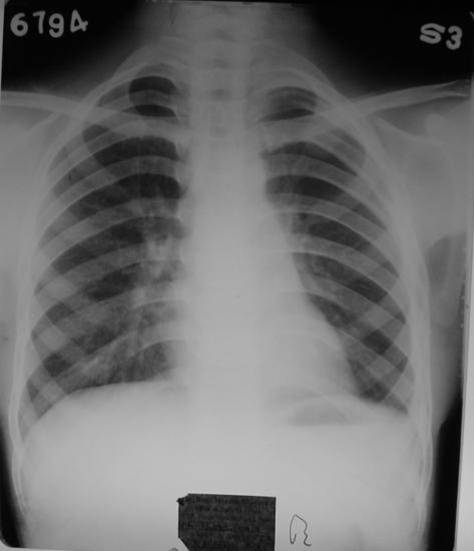
Collapsed lung with Empyema


This case improved gradually and gets cured within six months. Till date he has no problem .
Dated. 5 Dec.1992.
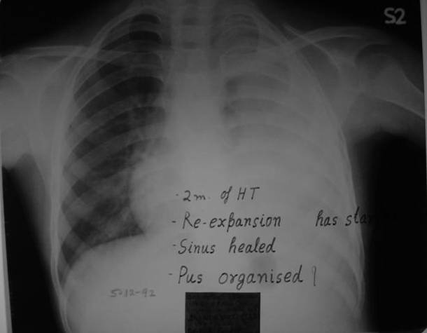
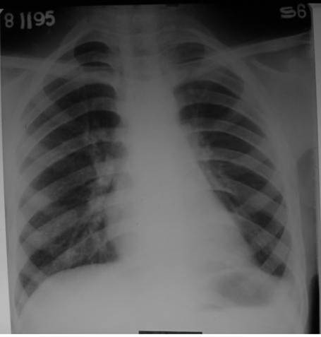
Re-expansion had started after one month of homeopathic treatment.
Dated: 8 Feb.1993.
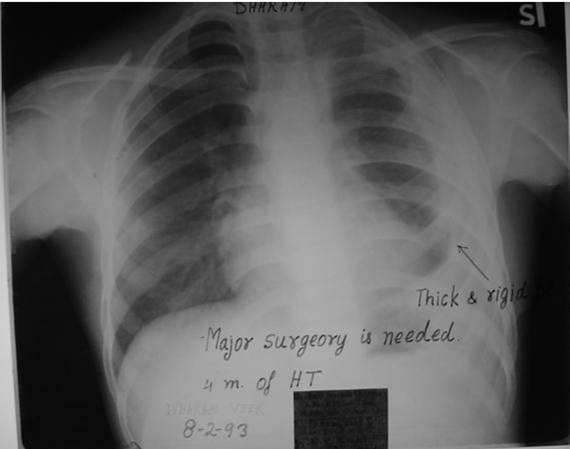
Re-expansion of collapsed lung was in progress. Sinus had healed.
Dated: 1 April 1993.
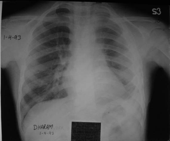
See the gradual non-stop re-expansion of lung. Pleura had become thick and fibrosed.
Dated: 6 Jan. 1994.
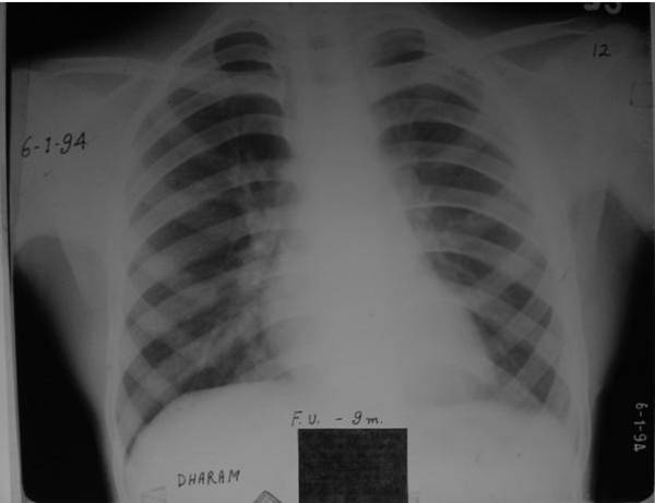
Again there is a process of resolution of fibrosed pleura is visible.
Discussion:
Pus in long standing Empyema becomes organized and fibrosed leads to
immobilization of the affected side of lung. As per Medical Literature
Fibrosis can not be reverted but it is not so in this case. Fibrosed
and thickened pleura ultimately gets normal in its shape.

Contact Form
We have been treating kidney problems with the help of many homeopathic doctors working with us in a team system. We are doing classical homeopathy (which is best of all) that is based on pure homeopathic principles that is using single medicine at a time and without homeopathic mixers or lequids or mother tinctures
Read More